Review


mouse pre myoblast cell line c2c12  (ATCC)


Bioz Verified Symbol ATCC is a verified supplier
Bioz Manufacturer Symbol ATCC manufactures this product  
  • Logo
  • About
  • News
  • Press Release
  • Team
  • Advisors
  • Partners
  • Contact
  • Bioz Stars
  • Bioz vStars
  • 99

    Structured Review

    ATCC mouse pre myoblast cell line c2c12
    Mouse Pre Myoblast Cell Line C2c12, supplied by ATCC, used in various techniques. Bioz Stars score: 99/100, based on 8447 PubMed citations. ZERO BIAS - scores, article reviews, protocol conditions and more
    https://www.bioz.com/product/mouse+pre+myoblast+cell+line+c2c12/10__3390_slash_polym18101184-79-19-24?v=ATCC
    Average 99 stars, based on 8447 article reviews
    mouse pre myoblast cell line c2c12 - by Bioz Stars, 2026-07
    99/100 stars
    Images


    Similar Products

    99
    ATCC mouse pre myoblast cell line c2c12
    Mouse Pre Myoblast Cell Line C2c12, supplied by ATCC, used in various techniques. Bioz Stars score: 99/100, based on 1 PubMed citations. ZERO BIAS - scores, article reviews, protocol conditions and more
    https://www.bioz.com/product/mouse+pre+myoblast+cell+line+c2c12/10__3390_slash_polym18101184-79-19-24?v=ATCC
    Average 99 stars, based on 1 article reviews
    mouse pre myoblast cell line c2c12 - by Bioz Stars, 2026-07
    99/100 stars
      Buy from Supplier

    99
    ATCC c2c12 mouse pre myoblast cell line
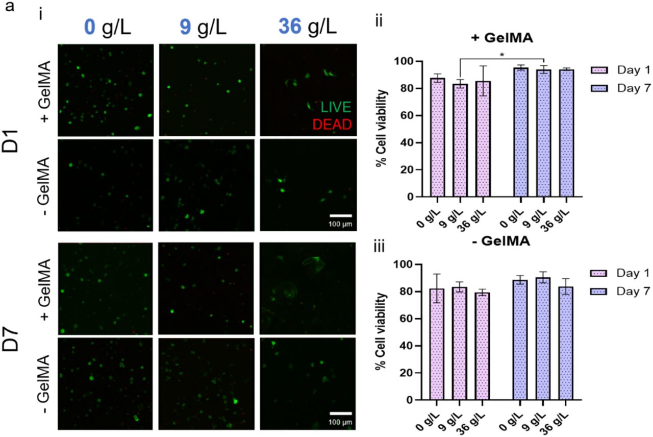
    Cell viability of <t>C2C12</t> in ATPS systems. a-i) Confocal images with C2C12 labelled with Calcein to show live cells (green) and cells labelled with propidium iodide to show dead cells (red). Quantification of cell viability in ATPS systems where a-ii) GelMA was cross-linked and in samples where a-iii) GelMA was removed. Scale bars: (a) 100 µm. Statistical significances were assessed by two-way ANOVA. Mean ± S.D. n=3, *p<0.05.
    C2c12 Mouse Pre Myoblast Cell Line, supplied by ATCC, used in various techniques. Bioz Stars score: 99/100, based on 1 PubMed citations. ZERO BIAS - scores, article reviews, protocol conditions and more
    https://www.bioz.com/product/mouse+pre+myoblast+cell+line+c2c12/bio_rxiv__2025__06__27__661968-296-8-22?v=ATCC
    Average 99 stars, based on 1 article reviews
    c2c12 mouse pre myoblast cell line - by Bioz Stars, 2026-07
    99/100 stars
      Buy from Supplier

    99
    ATCC mouse pre myoblast cell line
    Cell viability of <t>C2C12</t> in ATPS systems. a-i) Confocal images with C2C12 labelled with Calcein to show live cells (green) and cells labelled with propidium iodide to show dead cells (red). Quantification of cell viability in ATPS systems where a-ii) GelMA was cross-linked and in samples where a-iii) GelMA was removed. Scale bars: (a) 100 µm. Statistical significances were assessed by two-way ANOVA. Mean ± S.D. n=3, *p<0.05.
    Mouse Pre Myoblast Cell Line, supplied by ATCC, used in various techniques. Bioz Stars score: 99/100, based on 1 PubMed citations. ZERO BIAS - scores, article reviews, protocol conditions and more
    https://www.bioz.com/product/mouse+pre+myoblast+cell+line+c2c12/pm34996237-119-9-17?v=ATCC
    Average 99 stars, based on 1 article reviews
    mouse pre myoblast cell line - by Bioz Stars, 2026-07
    99/100 stars
      Buy from Supplier

    99
    ATCC pre myoblastic mouse cell line c2c12
    (A) Proportional Venn diagram. Fractions of called SNVs identical to the Genomes Project data and dbSNP130. Only data for which the minor allele frequency or the average heterozygosity was known and below 1% were used for comparison. (B) Distribution of synonymous, missense, nonsense and mutations affecting the start or stop codon are shown in relation to all somatic mutations. (C) BMPR1A mutations p.W487R and p.E502G are located at the protein kinase domain of BMPR1A. Reference amino acids are in green, the mutated forms are shown in red. The net structure at the left lower side indicates the ATP binding domain. (D) BMPR1A mutations show decreased signaling acitivity. Activity of wt mBMPR1A, mBMPR1A E502G and mBMPR1A W487R was determined in <t>C2C12</t> cells using a SMAD-responsive Luciferase reporter gene assay. Induced Luciferase activity was normalized to Renilla acitivty. The activity of untransfected cells was set to 0% and the activity of wt mBmpr1a was set to 100%. Significant differences were calculated with a two-tailed t-test and marked as: * p≤0.05, ** p≤0.01, *** p≤0.001.
    Pre Myoblastic Mouse Cell Line C2c12, supplied by ATCC, used in various techniques. Bioz Stars score: 99/100, based on 1 PubMed citations. ZERO BIAS - scores, article reviews, protocol conditions and more
    https://www.bioz.com/product/mouse+pre+myoblast+cell+line+c2c12/pmc03008745-195-22-27?v=ATCC
    Average 99 stars, based on 1 article reviews
    pre myoblastic mouse cell line c2c12 - by Bioz Stars, 2026-07
    99/100 stars
      Buy from Supplier

    Image Search Results


    Cell viability of C2C12 in ATPS systems. a-i) Confocal images with C2C12 labelled with Calcein to show live cells (green) and cells labelled with propidium iodide to show dead cells (red). Quantification of cell viability in ATPS systems where a-ii) GelMA was cross-linked and in samples where a-iii) GelMA was removed. Scale bars: (a) 100 µm. Statistical significances were assessed by two-way ANOVA. Mean ± S.D. n=3, *p<0.05.

    Journal: bioRxiv

    Article Title: Aqueous two-phase bioinks for discrete packing and compartmentalisation of 3D bioprinted cells

    doi: 10.1101/2025.06.27.661968

    Figure Lengend Snippet: Cell viability of C2C12 in ATPS systems. a-i) Confocal images with C2C12 labelled with Calcein to show live cells (green) and cells labelled with propidium iodide to show dead cells (red). Quantification of cell viability in ATPS systems where a-ii) GelMA was cross-linked and in samples where a-iii) GelMA was removed. Scale bars: (a) 100 µm. Statistical significances were assessed by two-way ANOVA. Mean ± S.D. n=3, *p<0.05.

    Article Snippet: The A549 human lung carcinoma epithelial-like cell line, C2C12 mouse pre-myoblast cell line and MG63 human osteosarcoma cell line were obtained from ATCC (USA).

    Techniques:

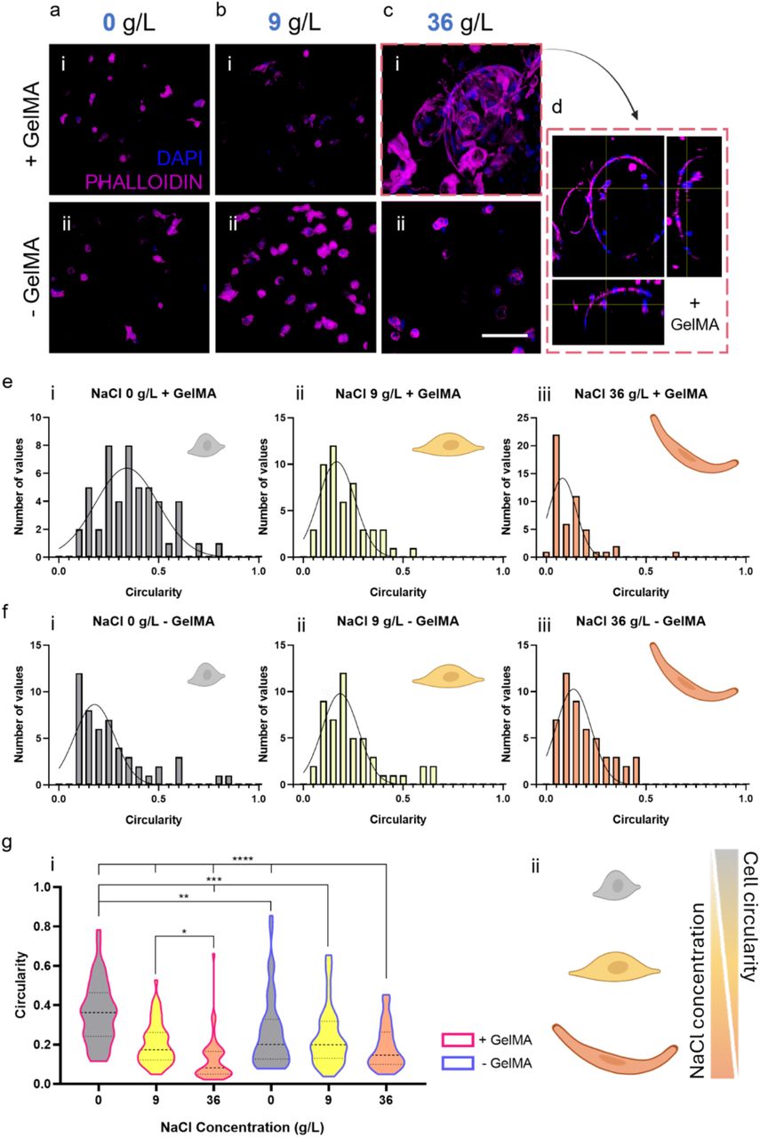
    Spatial distribution of C2C12 in ATPS systems. a-b-c) Confocal images with C2C12 labelled with DAPI to show nuclei (blue) and cells marked with phalloidin to show f-actin (magenta). d) Orthogonal view of image c-i. Circularity analysis conducted on the formulation e-i) NaCl 0 g/L with GelMA, e-ii) NaCl 9 g/L with GelMA, e-iii) NaCl 36 g/L with GelMA, f-i) NaCl 0 g/L without GelMA, f-ii) NaCl 9 g/L without GelMA and f-iii) NaCl 36 g/L without GelMA. g) Cell circularity as a function of salt concentration. g-i) Cell circularity for each NaCl composition within the ATPS samples is shown. The pink contour marks samples with + GelMA; the blue contour marks samples with - GelMA. g-ii) Cell circularity is inversely proportional to the salt concentration in the hydrogels. When the amount of NaCl in the scaffolds is higher, the cells are more elongated. Scale bars: (c) 100 µm. Statistical significances were assessed by two-way ANOVA. Mean ± S.D. n=3, ****p<0.0001, ***p<0.001, **p<0.01, *p<0.05.

    Journal: bioRxiv

    Article Title: Aqueous two-phase bioinks for discrete packing and compartmentalisation of 3D bioprinted cells

    doi: 10.1101/2025.06.27.661968

    Figure Lengend Snippet: Spatial distribution of C2C12 in ATPS systems. a-b-c) Confocal images with C2C12 labelled with DAPI to show nuclei (blue) and cells marked with phalloidin to show f-actin (magenta). d) Orthogonal view of image c-i. Circularity analysis conducted on the formulation e-i) NaCl 0 g/L with GelMA, e-ii) NaCl 9 g/L with GelMA, e-iii) NaCl 36 g/L with GelMA, f-i) NaCl 0 g/L without GelMA, f-ii) NaCl 9 g/L without GelMA and f-iii) NaCl 36 g/L without GelMA. g) Cell circularity as a function of salt concentration. g-i) Cell circularity for each NaCl composition within the ATPS samples is shown. The pink contour marks samples with + GelMA; the blue contour marks samples with - GelMA. g-ii) Cell circularity is inversely proportional to the salt concentration in the hydrogels. When the amount of NaCl in the scaffolds is higher, the cells are more elongated. Scale bars: (c) 100 µm. Statistical significances were assessed by two-way ANOVA. Mean ± S.D. n=3, ****p<0.0001, ***p<0.001, **p<0.01, *p<0.05.

    Article Snippet: The A549 human lung carcinoma epithelial-like cell line, C2C12 mouse pre-myoblast cell line and MG63 human osteosarcoma cell line were obtained from ATCC (USA).

    Techniques: Formulation, Concentration Assay

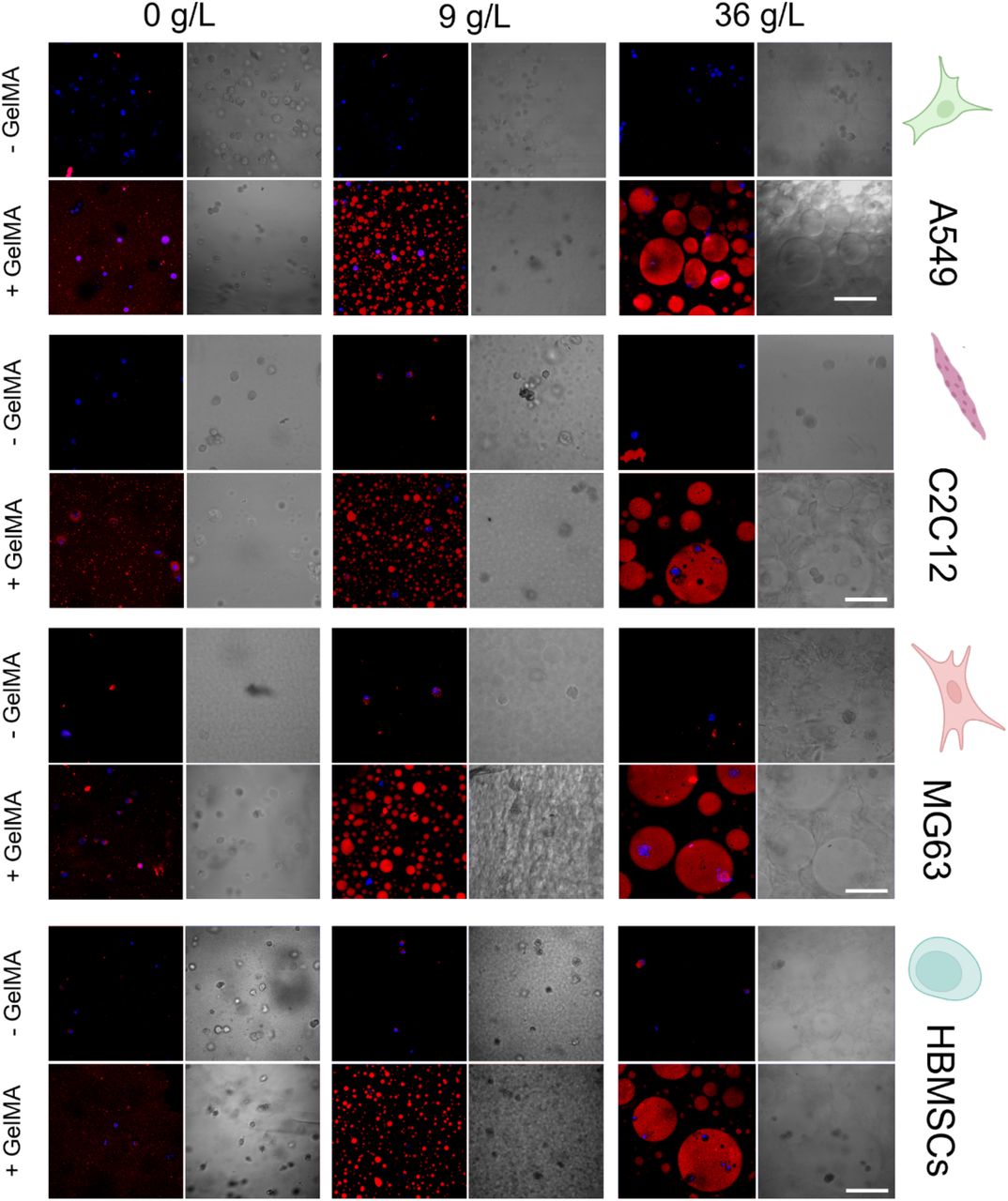
    Partitioning of different cell types within the various ATPS scaffold formulations. Brightfield and confocal (merged) images representative of cells labelled with DAPI to show the nucleus (blue) of the different cell types (A549, C2C12, MG63, HBMSCs), and GelMA marked with rhodamine B to show the inner phase (red) of the scaffolds at 0 - 9 - 36 g/L. In all cell types, images were taken both under conditions where GelMA was not chemically cross-linked (-GelMA) and under conditions where GelMA was chemically cross-linked (+GelMA). Scale bars: (a, b, c, d) 100 μm. Mean ± S.D. n=3.

    Journal: bioRxiv

    Article Title: Aqueous two-phase bioinks for discrete packing and compartmentalisation of 3D bioprinted cells

    doi: 10.1101/2025.06.27.661968

    Figure Lengend Snippet: Partitioning of different cell types within the various ATPS scaffold formulations. Brightfield and confocal (merged) images representative of cells labelled with DAPI to show the nucleus (blue) of the different cell types (A549, C2C12, MG63, HBMSCs), and GelMA marked with rhodamine B to show the inner phase (red) of the scaffolds at 0 - 9 - 36 g/L. In all cell types, images were taken both under conditions where GelMA was not chemically cross-linked (-GelMA) and under conditions where GelMA was chemically cross-linked (+GelMA). Scale bars: (a, b, c, d) 100 μm. Mean ± S.D. n=3.

    Article Snippet: The A549 human lung carcinoma epithelial-like cell line, C2C12 mouse pre-myoblast cell line and MG63 human osteosarcoma cell line were obtained from ATCC (USA).

    Techniques:

    (A) Proportional Venn diagram. Fractions of called SNVs identical to the Genomes Project data and dbSNP130. Only data for which the minor allele frequency or the average heterozygosity was known and below 1% were used for comparison. (B) Distribution of synonymous, missense, nonsense and mutations affecting the start or stop codon are shown in relation to all somatic mutations. (C) BMPR1A mutations p.W487R and p.E502G are located at the protein kinase domain of BMPR1A. Reference amino acids are in green, the mutated forms are shown in red. The net structure at the left lower side indicates the ATP binding domain. (D) BMPR1A mutations show decreased signaling acitivity. Activity of wt mBMPR1A, mBMPR1A E502G and mBMPR1A W487R was determined in C2C12 cells using a SMAD-responsive Luciferase reporter gene assay. Induced Luciferase activity was normalized to Renilla acitivty. The activity of untransfected cells was set to 0% and the activity of wt mBmpr1a was set to 100%. Significant differences were calculated with a two-tailed t-test and marked as: * p≤0.05, ** p≤0.01, *** p≤0.001.

    Journal: PLoS ONE

    Article Title: Somatic Mutation Profiles of MSI and MSS Colorectal Cancer Identified by Whole Exome Next Generation Sequencing and Bioinformatics Analysis

    doi: 10.1371/journal.pone.0015661

    Figure Lengend Snippet: (A) Proportional Venn diagram. Fractions of called SNVs identical to the Genomes Project data and dbSNP130. Only data for which the minor allele frequency or the average heterozygosity was known and below 1% were used for comparison. (B) Distribution of synonymous, missense, nonsense and mutations affecting the start or stop codon are shown in relation to all somatic mutations. (C) BMPR1A mutations p.W487R and p.E502G are located at the protein kinase domain of BMPR1A. Reference amino acids are in green, the mutated forms are shown in red. The net structure at the left lower side indicates the ATP binding domain. (D) BMPR1A mutations show decreased signaling acitivity. Activity of wt mBMPR1A, mBMPR1A E502G and mBMPR1A W487R was determined in C2C12 cells using a SMAD-responsive Luciferase reporter gene assay. Induced Luciferase activity was normalized to Renilla acitivty. The activity of untransfected cells was set to 0% and the activity of wt mBmpr1a was set to 100%. Significant differences were calculated with a two-tailed t-test and marked as: * p≤0.05, ** p≤0.01, *** p≤0.001.

    Article Snippet: The activity of the wildtype mouse protein (wtmBMPR1A) and its mutants was determined by measuring induced Luciferase activity in the transiently transfected pre-myoblastic mouse cell line C2C12 (ATCC).

    Techniques: Comparison, Binding Assay, Activity Assay, Luciferase, Reporter Gene Assay, Two Tailed Test

